- Have any questions? Contact us!
- info@dr-rath-foundation.org


President Obama, Wake Up America!
November 13, 2010
Brussels EU continues attempts to curtail spread of lifesaving natural health information
May 4, 2011How to use diagnostic technologies wisely and for the benefit of your health
By Bilwa Bhanap, MD, Aleksandra Niedzwiecki, PhD
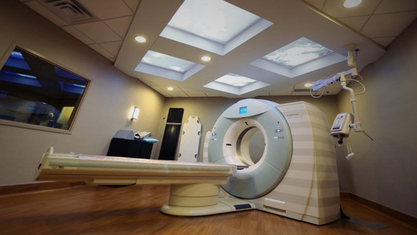
The early detection of health problems is very important in curtailing the progress of, or eliminating, disease at its onset. Over the last decades, various imaging techniques such as X-rays, ultrasounds, MRI and CT scans have been developed and applied to diagnostic, as well as therapeutic, medical care.
However, in recent years, many doctors – and especially radiologists – have become concerned with the overuse of certain diagnostic techniques, in particular those that expose patients to radiation. Although infrequent use of X-ray or CT (computed tomography) scans will not have adverse effects on a patient, multiple exposures to radiation over a short period of time can cause serious damage to cells, resulting in an increased risk of cancer and other diseases.
In addition to illustrating some of the concerns associated with medical radiation exposure, the following overview presents ways to protect our bodies and minimize the risks of damage.
Sources of radiation exposure
Our bodies are exposed to different types of radiation coming from a variety of sources, such as non-ionizing radiation from microwaves, electric power lines or radio signals, and a natural ionizing radiation originating from substances contained in the earth’s crust and space. In medicine, many diagnostic imaging technologies, such as X-ray machines and CT scans, were designed to look for abnormalities inside of the body’s organs and are based on radiation.
These medical diagnostic technologies are increasingly becoming more popular. The use of CT scans in the US has skyrocketed over the past three decades, from 3 million scans in the 1980s to 70 million scans in 2007. This rate has doubled in the last 5 years. More than 4-7 million children are getting CT scans, and the numbers continue to increase by 10% every year.
Increasing health concerns with the use of radiation-based diagnostics
The controversy about CT scans being an increasing source of radiation exposure – and thus increasing the risks of future cancers – caught public attention in 2007. In a study published in the New England Journal of Medicine, Dr. David Brenner, the lead author, highlighted various concerns related to a large increase in the use of CT scans in children and adults. He emphasized that only a small percentage of scans are absolutely necessary and that about one-third can be replaced by other approaches or not done at all. Another study published in the same year, sponsored by the National Institutes of Health (NIH) and the National Cancer Institute (NCI), estimated that the increased exposure may lead to 29,000 new cases of cancers in upcoming years.
Despite the continually increasing popularity of CT scans, there are very few regulations regarding their use. In a study published in 2009 by the University of California in San Francisco (UCSF), researchers found that there is up to 13-fold variation in the radiation doses for similar tests conducted using different machines or settings, sometimes even at the same medical facility. In 2009, a major hospital came under FDA investigation for over-exposing more than 200 patients due to improper settings in brain scans. Similar issues were revealed at other centers in the USA.
Why are so many CT scans being performed?
One of the reasons for the increase in CT scans is that they are a non-invasive, fast and painless testing method with which to identify tissue abnormalities and developing health problems. Many CT scans provide high resolution 3-D images, which make it easier to view details of internal organs, blood vessels, bones and tumors. Also, as the scan can be taken within seconds, CT scan images are easy to take – as opposed to X-Rays, ultrasounds, and MRIs, which require the patient to remain still for long period of time.
The popularity of CT scans also relates to the fact that many physicians use multiple testing as a protection against malpractice claims. Some physicians, when unsure as to which is the best test to order for a particular symptom, often choose a CT scan so as to cover a wide range of potential problems. However, sometimes for appendicitis in a child, an ultrasound exam is better and far less dangerous than a CT scan. Quite often, multiple CT images are taken in one setting to give a clearer idea of a potential problem.
Cost can also play a role because the insurance “fee-for-procedure” method rewards doctors for doing more tests. Many commercial centers promote annual testing to screen the whole body for early detection of diseases. Their customers, who are sometimes referred to as the “worried well,” pay out-of-pocket expenses to get that extra dose of radiation. Many patients request this test because another friend with similar symptoms had also had a CT scan.
In many cases, the over-use of radiation-based technologies relates to a lack of awareness about the associated health risks. According to a 2003 survey, approximately 75% of radiologists and emergency-room physicians significantly underestimate the radiation dose from CT scans. They seem to think that the radiation exposure through CT is equal or marginally more than a conventional X-Ray. In fact, the radiation from one CT scan can be equivalent to approximately 500 chest X-Rays, with a wide range of variation depending upon the body area scanned. For instance, a cardiac CT scan recording calcium deposits has a much lower dose of radiation exposure (equal to one mammogram scan) than a CT angiogram, the latter of which exposes a patient to multiple higher doses (the equivalent of 20 mammogram scans).
Some orthodontists, especially those with the highest percentage of pediatric patients, say they feel “blind-folded” if working without a cone-beam CT scan. Some also use the “fun-factor” of viewing 3-D skull images as a selling point for their practice. The scan technologists tend to increase the radiation per dose to get “amazing” pictures with more contrast.
Following recent regulations, the US Transportation Security Administration will be scanning all airline passengers with X-ray scanners, thus further exposing millions of travellers to radiation. Although an individual’s lifetime risk of developing cancer due to radiation exposure may not be high, with millions of scans being performed every year, the cumulative risk for the population is much higher. As such, this is a huge public health issue and potential cancer epidemic.
Who is at risk?
In general, there is no “safe” level of radiation exposure. In particular, children, young adults and women have the highest risk of developing radiation-induced cancer in their lifetime.
Children and young adults: Children are 10 times more sensitive to the same dose of radiation than an adult. Nevertheless, an increasing number of children are getting CT scans because it is convenient for the doctor and parents. Approximately one-third of children getting a CT scan are 10 or younger. Yet, the machine settings are rarely changed for a child’s CT scan. There are many concerns with radiation-based tests conducted in children, which include:
- A child’s torso is thin, thus providing less protection from radiation-induced damage.
- Children have a longer life span to manifest the hazards of radiation.
- Immature and rapidly dividing cells (i.e. blood cells) and developing organ systems are more prone to DNA damage from radiation exposure, which increases a risk of developing organ abnormalities or diseases.
According to Dr. Brenner’s 2007 study, an estimated 33% of CT scans are unnecessary. This translates into more than 1.5-2 million children getting very high doses of radiation that has detrimental health effects
Women: A UCSF researcher, R. Smith-Bindman, projected that 1 in 270 women aged 40 who undergo a cardiac angiography CT scan to study coronary blood vessels will develop some form of cancer, as opposed to 1 in 600 men of the same age who undergo the same procedure.
Although all body organs could potentially be harmed by radiation exposure, the breast, thyroid, bone marrow, digestive and reproductive organs are more radiosensitive due to continuous cellular growth activity. It is reported that a higher radiation dose is used for head scans, yet the risk of potential harm is higher in abdominal scans due to organ sensitivity.
The role of the radiologist as a gatekeeper
In a typical scenario, a patient is referred by a physician to a radiologist for a specific scan. A technician performs the scan, prepares the images and preliminary results for the radiologist to review and sign and the office sends out the bill. Special consideration is rarely given to any other aspects of the patient’s history, symptoms, possible diagnosis, age, height and weight, or whether there is a better option than performing a CT scan.
Increased usage of CT scans does not add value to patients’ medical care, but does put them – and future generations – on a trajectory for cancer, heart disease and genetic malformations.
In a 2010 meeting of the Radiological Society of North America, in a campaign named “Image Wisely,” more than 700 radiologists pledged to use less radiation on their patients. This campaign aims to better educate health practitioners and patients to keep radiation exposure to a minimum level. The International Commission on Radiological Protection already recommends using the ALARA (“as low as reasonably achievable”) principle for limiting the radiation doses received by people. The Image Wisely campaign is based on a previously successful “Image Gently” campaign launched in 2008. The Image Gently campaign encourages the usage of imaging protocols specifically modified for children. These two campaigns mainly focus on standardization of protocols, reducing multi-scan images, and the optimization of radiation exposure based on each individual patient. At present, however, use of these recommendations remains voluntary and the efforts are still in their infancy. It is therefore up to the individual to take control of his/her radiation exposure and, thereby, their health.
What a patient can do to minimize radiation exposure
The best defense is knowledge and a dose of healthy skepticism. You need to be an advocate for yourself and your children. There are some steps that you can take right away:
- Avoid repetitive tests: Keep your own accurate records of exposure to X-rays and CT scans, along with the reports. Patients with previous emergency (ER) visits for chest pain, kidney stones, and abdominal pain are more likely to have multiple tests.
- Go to facilities committed to radiation safety. Some institutions follow the American College of Radiology accredited protocols, with staff trainings on appropriate machines. However, such certification is currently not mandatory for CT scan owners.
- Ask questions:
- What information is the doctor looking for when a CT scan is suggested?
- How will this information improve the diagnosis/treatment/outcome of the disease?
- Are there any alternatives through which the same information can be obtained?For example, cardiac Ultrafast CT exposes the body to less radiation than the angiogram CT. An MRI scan is equally effective to provide images of the liver, pancreas and kidneys. As far as possible, parents may suggest using ultrasound tests for children as a substitute for radiation-based tests. The application of the ALARA (“as low as reasonably achievable”) principle is advised for ultrasound tests as well. Will the radiation dose be adjusted according to the size of the person? The dose should be much lower for children and adjusted for adults based upon their body mass index (BMI).
- Can the scan range be limited to a specific region only?Reduce exposure to multiphasic scans with contrast (e.g., 64 sliced scans are popular for cardiac studies).
- Consider financial interests, such as owning a machine.Many cardiologists, oncologists and some primary physicians purchase CT machines to increase their revenue sources. With inadequate staff training on appropriate radiation doses, higher doses are given to get clearer images. Some states like California, New York, Illinois have prohibited physician self-referral for CT scans.
Use of CT scans for screening
Use of radiation-based tests is important for the diagnosis and monitoring of high risk patients. However, the concern is greater when tests are performed on healthy populations for information purposes.
Whole body CT scans: Health-conscious people often want to get a whole body CT scan in the hope that it will aid the early detection of diseases. In addition to the extra radiation that such scans subject the body to, an additional issue with such screening is that it can generate suspicious results which may eventually turn out to be harmless. Yet, in the meantime, until given the “all-clear,” the person undergoes many invasive procedures and a lot of anxiety.
Mammography: While mammograms have helped in the detection and treatment of many cases of breast cancer, it is important that women talk to their physicians about the requirements and frequency of their mammogram tests. Mammogram radiation exposure can vary and can sometimes be equal to 75 chest X-rays. Considering the risk-benefit ratio and cost, regular yearly mammograms in women over 40 may not be essential and, especially, not necessary for every woman. A comparative study in breast cancer detection between the USA and UK found although the cancer detection rates were similar in both countries, false positive test results were twice as high in the US. To save patients a lot of psychological and financial burden, it is important that mammograms are evaluated by an experienced physician.
Lung Cancer screening: Preliminary findings of a government funded study presented in November 2010 indicated that annual CT scans of current and former smokers may reduce the death risk from lung cancer and other causes. As a result, without a thorough peer-review assessment and confirmation of the findings of this study, many commercial screening centers are already advertising such CT scans for a much broader population, including nonsmokers. Similar to other screening scans, in addition to unnecessary radiation, there is a high possibility of detecting false positives or benign abnormalities. Such screening, followed by the “treatment” of diseases that never existed, could prove more harmful than useful.
It is very important to diagnose and prevent diseases using appropriate advances in medical technology. However, the benefits of radiation-based scans should always be weighed against the risk of radiation damage.
Consequences of radiation damage to our body cells
The damaging effects of ionizing radiation on cells can be summarized as follows:
- Ionizing radiation can cause damage in two ways:
- Directly: Radiation damages cellular macromolecules, e.g., DNA and proteins leading to DNA mutation and cell death
- Indirectly: Radiation interacts with water molecules in the cell, generating free radical unstable hydrogen peroxide (H2O2) which has a toxic effect on cells and can cause cell damage or even death.
- Radiation-induced carcinogenesis can occur in two ways:
- Initiation effect: Radiation can cause a genetic mutation that can convert normal, rapidly developing, cells into precancerous cells leading to cancers. Children and young adults are more susceptible to this effect.
- Promotion effect: This effect could be more active in adults who already have pre-existing cancerous cells. External radiation prompts rapid growth and development of these cells.
How to protect the body’s cells from unavoidable radiation exposure
Incorporate a synergistic nutrient program in the daily regimen and adjust it before the CT scan. Nutrients, especially antioxidants such as vitamins C, A, and E, green tea extract, quercetin and others are reported to prevent the formation of oxygen free radicals and reduce the toxicity of already formed free radicals, thereby avoiding cellular damage in multiple ways.
Vitamins C, A and E – In addition to other protective effects, vitamin C is most important in preventing genetic damage to chromosomes. In addition, it also protects bone marrow cells and acts synergistically with antioxidants such as vitamins A and E to prevent the radiation-induced death of healthy cells and induce cell death (apoptosis) in damaged cells.
Phytonutrients:
- Green tea extract is unique in providing protection to rapidly dividing cells in the digestive system and bone marrow and in reducing the damaging effects of radiation impact.
- Quercetin provides protection to chromosomes and mitochondrial DNA.
- Resveratrol protects the most radio-sensitive tissues and organs, such as the liver and the digestive tract, from radiation damage. In addition, it also has selective action on cancer cells.
- Curcumin protects normal cells from radiation damage, as well as increasing the activity of the genes responsible for cancer cell death.
Other nutrients:
- N-Acetyl-Cysteine (NAC) is known to synergistically act with vitamins C and E to protect the cells, even when taken immediately after radiation exposure.
- Zinc and Manganese are the cofactors of natural antioxidant enzymes which are important components of a cellular anti-oxidant defense system. Zinc is known to protect bone marrow – and especially red blood cells – against damage, but not tumor cells. Zinc and Manganese together are powerful in protecting cellular mitochondria, which fuel our cells with bio-energy.
Although many of these studies are conducted in research laboratories, it is already recommended that airline pilots, other crews and astronauts take high doses of antioxidant nutrients. However, due to lack of awareness, physicians do not recommend the same to their patients.
While the guidelines are still blurry on who should or should not get CT scans, and what their exact indications and frequency should be, our knowledge and open discussion with the physician are the keys for minimizing the health risks of these diagnostic tests. In addition, it is very important to ensure an optimum daily intake of antioxidant nutrients and increase it as necessary.
References:
- Brenner D, Hall E. Computed Tomography: An Increasing Source of Radiation Exposure. New England Journal of Medicine 2007; 357:2277-84
- Smith-Bindman R, Lipson J, Marcus R, Kim KP, Mahesh M, Gould R, Berrington de González A, Miglioretti DL. Radiation dose associated with common computed tomography examinations and the associated lifetime attributable risk of cancer. Arch Intern Med. 2009 Dec 14;169(22):2078-86.
- Smith-Bindman R, Chu PW, Miglioretti DL, Sickles EA, Blanks R, Ballard-Barbash R, Bobo JK, Lee NC, Wallis MG, Patnick J, Kerlikowske K. Comparison of screening mammography in the United States and the United Kingdom. JAMA. 2003 Oct 22;290(16):2129-37.
- National Lung Screening Trial Research Team, Aberle DR, Adams AM, Berg CD, Clapp JD, Clingan KL, Gareen IF, Lynch DA, Marcus PM, Pinsky PF. Baseline characteristics of participants in the randomized national lung screening trial. J Natl Cancer Inst. 2010 Dec 1;102(23):1771-9. Epub 2010 Nov 22.
- Yong LC, Petersen MR, Sigurdson AJ, Sampson LA, Ward EM. High dietary antioxidant intakes are associated with decreased chromosome translocation frequency in airline pilots. Am J Clin Nutr. 2009 Nov;90(5):1402-10. Epub 2009 Sep 30.
- Shuryak I, Sachs RK, Brenner DJ. Cancer risks after radiation exposure in middle age. J Natl Cancer Inst. 2010 Nov 3;102(21):1628-36. Epub 2010 Oct 25.
- Brenner DJ. Slowing the increase in the population dose resulting from CT scans. Radiat Res. 2010 Dec;174(6):809-15. Epub 2010 Aug 23.
- Brenner DJ. Should we be concerned about the rapid increase in CT usage? Rev Environ Health. 2010 Jan-Mar;25(1):63-8.
- Okunieff P, Xu J, Hu D, Liu W, Zhang L, Morrow G, Pentland A, Ryan JL, Ding I. Curcumin protects against radiation-induced acute and chronic cutaneous toxicity in mice and decreases mRNA expression of inflammatory and fibrogenic cytokines. Int J Radiat Oncol Biol Phys. 2006 Jul 1;65(3):890-8.
- Velioğlu-Oğünç A, Sehirli O, Toklu HZ, Ozyurt H, Mayadağli A, Ekşioğlu-Demiralp E, Erzik C, Cetinel S, Yeğen BC, Sener G. Resveratrol protects against irradiation-induced hepatic and ileal damage via its anti-oxidative activity. Free Radic Res. 2009;43(11):1060-71.
- Tobi SE, Gilbert M, Paul N, McMillan TJ. The green tea polyphenol, epigallocatechin-3-gallate, protects against the oxidative cellular and genotoxic damage of UVA radiation. Int J Cancer. 2002 Dec 10;102(5):439-44.
